
Fully Digital X-ray Systems
Philips has a long history as a manufacturer of medical devices. The first Philips X-ray tubes were made as early as 1919. Today, Philips’ product range includes most of the imaging equipment needed in hospitals: magnetic resonance and computed tomography scanners, X-ray and fluoroscopy equipment, angiographs and fluoroscopes, ultrasound equipment and imaging workstations.
Although Philips still manufactures its X-ray tubes in-house, in Hamburg’s Röntgenstraße, innovation in the field of radiographs continues.
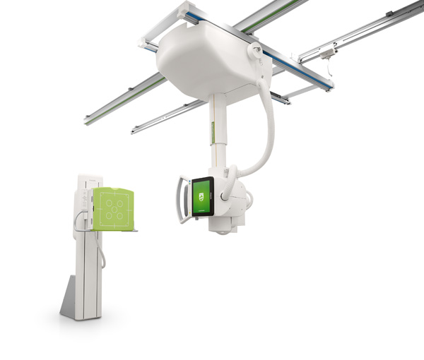
Philips Radiography 7300 C
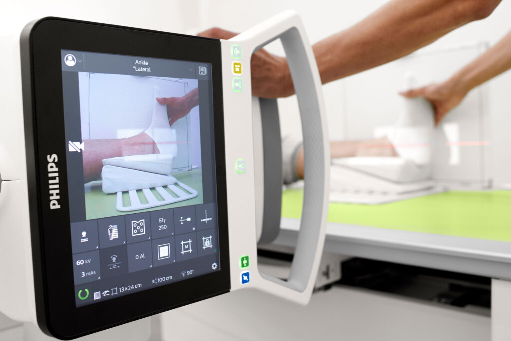
Philips’ state-of-the-art, AI-enabled, RedDot industrial design award-winning X-ray unit, the “Philips Radiography 7300 C”, is a user- and patient-friendly radiograph with low radiation dose and best image quality, which can be configured to meet the customer’s clinical needs.
- The intuitive software and hardware user interface enables new users to quickly begin operation.
- Unique “Variofocus” technology – shared use of focal spots ensures optimal image quality and the longest X-ray tube lifespan.
- AI options – 3D camera-based automatic collimation and AEC chamber indication.
- Many different configurations according to clinical need.
DigitalDiagnost C90
The Philips DigitalDiagnost C90 is designed for the acquisition, processing, storage, display, and export of digital radiographic images. The DigitalDiagnost C90 is suitable for all routine radiographic examinations, including specialties such as intensive care, traumatology and paediatrics, except fluoroscopy, angiography and mammography.
The system is suitable for all routine radiographic examinations, including specialties such as intensive care, traumatology and paediatrics. The system also uses more precise applications such as bone suppression.
Standard radiography procedures include:
• X-ray examinations of the skeleton, including the skull, chest, spine, pelvis, upper limbs, lower limbs, etc.;
• X-ray examinations of the lungs;
• X-ray examinations of soft tissue, e.g., the abdomen;
• full spine imaging in the patient’s vertical and horizontal positions;
• full leg imaging in the patient’s vertical and horizontal positions.
The following acquisition techniques are available to the user:
• detector acquisition with vertical, horizontal, and/or oblique X-ray beam;
• flexible acquisition technique for the flexible SkyPlate detector;
• flexible acquisition technique for Philips or other suppliers’ CR systems and film cassettes;
• manual acquisition techniques: kV-mA-ms; kV-mAs; kV-mAs-ms;
• automatic acquisition technique: AEC kV, mA, AEC-kV with falling load.
Device information leaflet.
Patient survey protocols:
You can see the range on the Philips website.